金桔
金币
威望
贡献
回帖0
精华
在线时间 小时
|
登陆有奖并可浏览互动!
您需要 登录 才可以下载或查看,没有账号?立即注册


×
高维多色流式
随着生物科技日新月异,越来越多检测高维单细胞免疫表型的技术脱颖而出,这其中包括:质谱流式,光谱流式,单细胞测序和多色高维流式等方法,这些方法都帮助我们以更多的参数揭示了对免疫系统复杂性的深刻见解,本篇小优以多色流式为例带大家看看研究高维免疫表型检的标准流程:
探究高维免疫分型实验整体流程如下图 1-6所示:
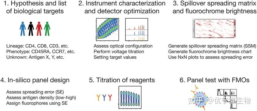
具体实验探究步骤
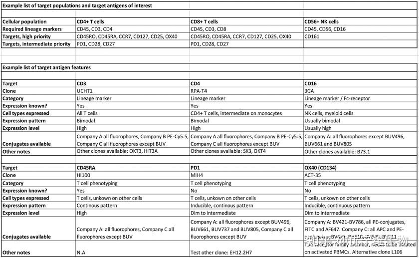
Step 1:选择、归类研究的免疫表型群体,如图2所示(以T细胞为例)
Step 2:电压滴定(了解流式仪器性能参数):
1:获得样本(常用PBMC或小鼠脾细胞)
2:选取仪器上所有通道的荧光抗体偶联特定蛋白(通常针对一个谱系标记,如CD4)
3:根据仪器指示执行流式细胞仪启动和每日QC。
4:记录每个单染样品,以30v的增量调整电压,从200v增加至700v,每调整一次电压变化 记录10000个细胞数量,保存并相应命名(例:BV421_200V, BV421_230V, BV421_260V等)
5:所有的单染样品依次重复词步骤,数据分析,计算 SI 值SI =MFI(pos) -MFI(neg)/(2 * rSD),得到的结果如下图3所示
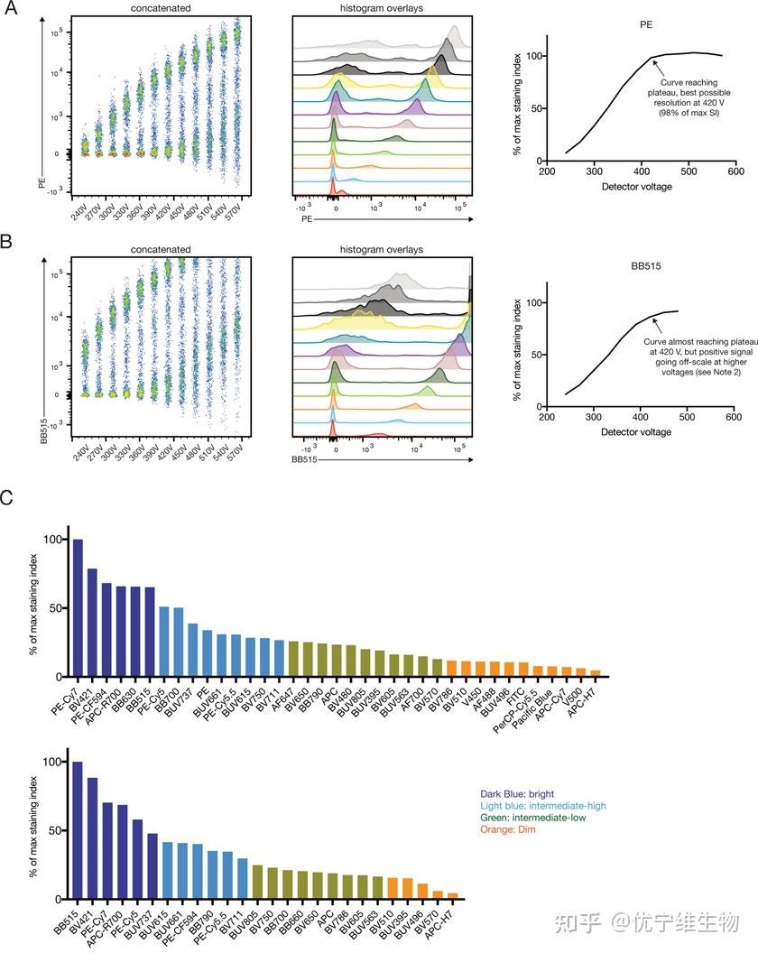
通过电压滴定我们可以很好的通过样本的分群情况来确认使用仪器的每个通道的最优电压,同时通过SI 值的计算 可以更精准的帮助我们了解使用仪器通道匹配荧光素的性能情况。
Step 3:补偿校准(了解仪器适配荧光素补偿情况)
每台流式细胞仪在捕捉特定荧光信号主峰和同时避免来自其他荧光团的信号之间保持平衡,但测量的不同荧光团之间仍会有光谱重叠(荧光素的光谱重叠、补偿和扩散误差如图4所示),无法避免的光谱重叠可以通过计算SE(spreading error 度量误差)来纠正,在Flowjo中我们可以使用 SSM(溢出扩散误差矩阵)帮助我们更好的了解到仪器各通道之间的SE情况。
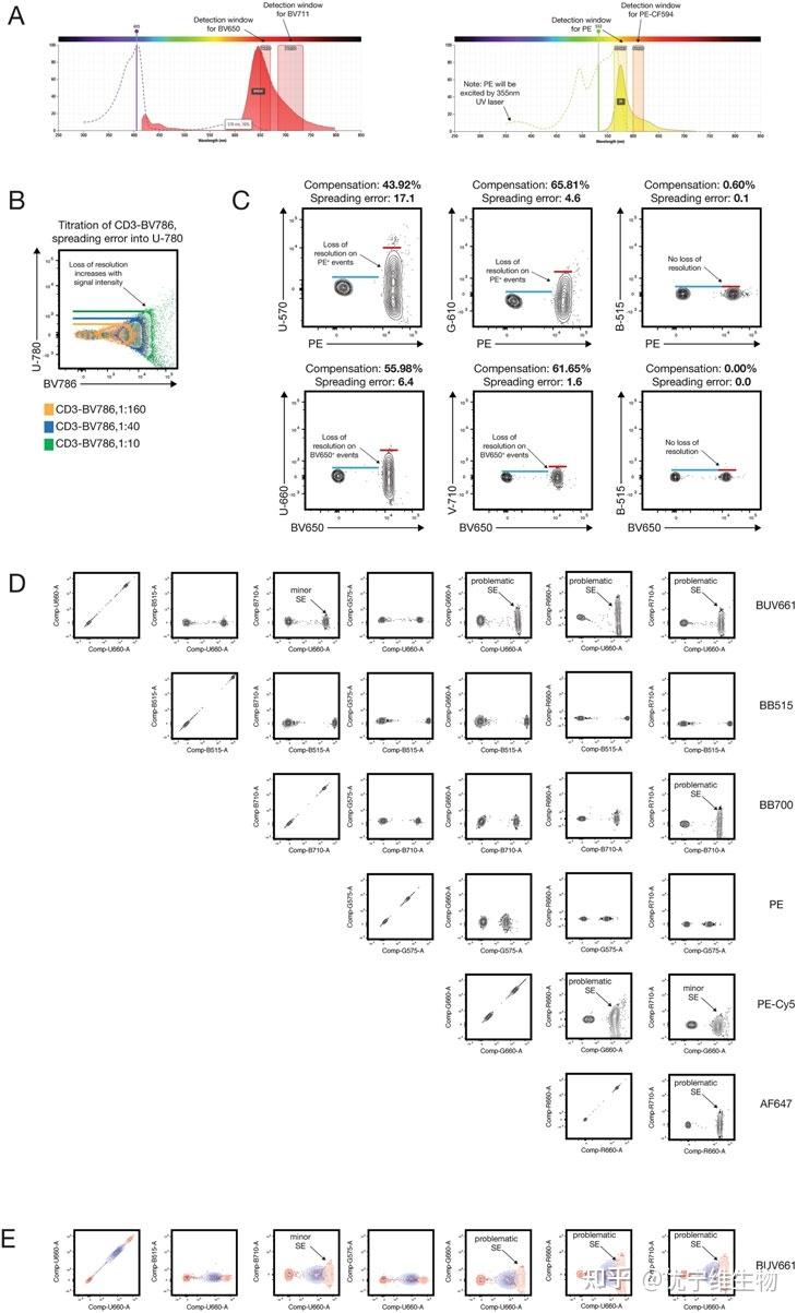
1:准备补偿微球,制备每个检测通道单染管。
2:通过step2 获取的最优电压情况,依次单染管上样,记录获取数据后,导入Flowjo,SSM 计算情况如下图5所示
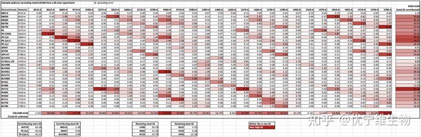
案例图示给出了由28色实验计算出的具有代表性的溢出扩散矩阵(SSM)。颜色从无SE(白色)到高SE(红色)从图中可以看出,最小SE的荧光团是BB515、AF647和BUV395。依次,接收最小SE的三个探测器是B-515, V-510和V-450,后续的panel设计中利用SSM 的数据可以最到限度避免补偿过大造成的结果偏差。
Step 4:panel 设计
在panel设计中最重要的2个因素:
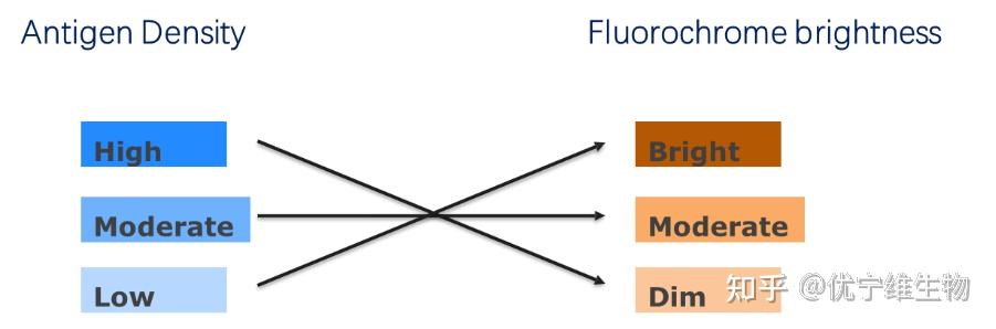
①:荧光素的亮度
流式panel设计的第一要素就是遵循“强弱”搭配原则,即:高表达的maker搭配亮度较暗的荧光素,低表达的maker搭配亮度较亮的荧光素,只有这种方式才可以平衡仪器通道中各类荧光素 且达到最优检测分群。如图6所示
② :补偿问题
流式panel设计的在遵循“强弱”搭配原则后考虑的第二要素,就荧光之间的补偿问题,通过Step3 我们已经获得SSM ,通过矩阵数据,我们可以轻松避免SE较大的2类荧光素标记共表达的maker。
遵循上述2个因素,设计panel ,选择对应抗体。
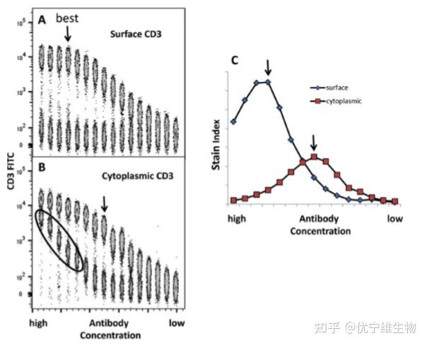
Step 5:抗体滴定
在选择试剂后,滴定是一个经常被忽略的重要步骤。滴定抗体的主要原因有两个:第一,减少非特异性结合的背景;第二,确保在饱和浓度下进行染色。对于典型的滴定实验,建议从每次染色反应的最高浓度2 μg开始进行8-12次连续稀释,选择最优抗体浓度。如图7所示
Step 6:Panel检测及对照设置
所有试剂都滴定完毕,就可以对整个panel进行测试,在整个实验中还必须去设置适当的对照(流式常用的对照:FMO、ISO等),通过对照可以评估荧光素之间可能因不可预见的spillover或相互作用而出现的问题,以FMO 为例 如图8所示:
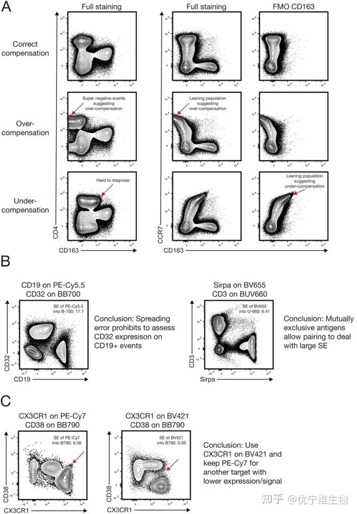
案例图示中利用 FMO 对照,对比 3种不同补偿状态样本的表达情况。
Step 7 数据分析
检测获得的FCS 格式文件由FlowJo (BD Biosciences),分析处理。
以上7个步骤是高维多色流式实验非常严谨的标准流程,有正在做的老师希望通过今天的分享可以帮助您的实验。
参考文献:
1. Robinson JP, Roederer M (2015) History ofscience flow cytometry strikes gold. Science350:739–740. https://doi.org/10.1126/science.aad6770
2. Spitzer MH, Nolan GP (2016) Mass cytometry: single cells, many features. Cell165:780–791. https://doi.org/10.1016/j.cell.2016.04.019
3. Zheng GXY, Terry JM, Belgrader P et al(2017) Massively parallel digital transcriptional
profiling of single cells. Nat Commun8:14049. https://doi.org/10.1038/ncomms14049
4. Brodie TM, Tosevski V (2017) Highdimensional single-cell analysis with mass cytometry. Curr Protoc Immunol 118:5.11.1–5.11.25. https://doi.org/10.1002/cpim.31
5. Papalexi E, Satija R (2017) Single-cell RNAsequencing to explore immune cell heterogeneity. Nat Rev Immunol 510:363. https://doi.org/10.1038/nri.2017.76
6. Mair F, Prlic M (2018) OMIP-044: 28-colorimmunophenotyping of the human dendritic
cell compartment. Cytometry A 106:255.https://doi.org/10.1002/cyto.a.23331
7. Futamura K, Sekino M, Hata A et al (2015) Novel full-spectral flow cytometry with multiple spectrally-adjacent fluorescent proteins and fluorochromes and visualization of in vivo cellular movement. Cytometry A 87:830–842. https://doi.org/10.1002/cyto.a.22725
8. Feher K, Volkmann von K, Kirsch J et al (2016) Multispectral flow cytometry: the consequences of increased light collection. Cytometry A 89:681–689. https://doi.org/10.1002/ cyto.a.22888
9. Kvistborg P, Gouttefangeas C, Aghaeepour N et al (2015) Thinking outside the gate: singlecell assessments in multiple dimensions. Immunity 42:591–592. https://doi.org/10.1016/j. immuni.2015.04.006
10. Saeys Y, Gassen SV, Lambrecht BN (2016) Computational flow cytometry: helping to make sense of high-dimensional immunology data. Nat Rev Immunol 16:449–462. https:// http://doi.org/10.1038/nri.2016.56
11. Mair F, Hartmann FJ, Mrdjen D et al (2016) The end of gating? An introduction to automated analysis of high dimensional cytometry data. Eur J Immunol 46:34–43. https://doi. org/10.1002/eji.201545774
12. Chester C, Maecker HT (2015) Algorithmic tools for mining high-dimensional cytometry data. J Immunol 195:773–779. https://doi. org/10.4049/jimmunol.1500633
13. Meinelt E, Reunanen M, Edinger M et al Standardizing application setup across multiple flow cytometers using BD FACSDiva™ Version 6 Software: technical bul
14.Ashhurst TM, Smith AL, King NJC (2017) High-dimensional fluorescence cytometry. Curr Protoc Immunol 10:5.8.1–5.8.38. https://doi.org/10.1002/cpim.37
15.Liechti T, Gu¨nthard HF, Trkola A (2018) OMIP-047: high-dimensional phenotypic characterization of B cells. Cytometry A 103:2262–2596. https://doi.org/10.1002/ cyto.a.23488
更多推荐:
原文地址:https://zhuanlan.zhihu.com/p/608068206 |
|

 /3
/3 

 浙公网安备33010802005999号


 2026庆【网站十三周
 2025庆【网站十二周
 2024庆中秋、迎国庆
 2024庆【网站十一周
 2023庆【网站十周年
 2022庆【网站九周年

 雷达卡
雷达卡 发表于 2025-5-28 07:02
 提升卡
提升卡